

Coccidiosis in Cattle
By Murray J. Kennedy, Food Safety Division, Alberta Agriculture and Food, Ropin The Web. Coccidiosis is a common parasitic protozoan disease of cattle, particularly weaned calves, in Alberta.Bovine coccidiosis is seen most frequently in calves that are three weeks to six months of age. Calves become infected when placed on pastures or lots contaminated by older cattle or other infected calves. Mature cattle may become infected when they are brought in from pastures and crowded into feedlots or barns.
At least nine species of coccidia occur in Alberta cattle, but only two, Eimeria zuernii and Eimeria bovis, cause severe clinical disease. To a lesser extent, Eimeria alabamensis also can cause clinical disease. The prevalence of the different species of coccidia can vary considerably between farms, regions, seasons and age groups.
Life Cycle of Coccidia
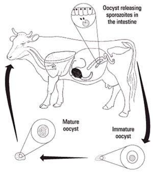
Bovine coccidia have stages both within the host animal as well as outside. The developmental stages in the animal give rise to a microscopic egg (called an oocyst), which is passed out in the manure (Figure 1).
Under proper conditions of temperature, moisture and oxygen, the oocyst develops within three to seven days and is now capable of infecting cattle. At this stage, the oocyst contains eight bodies (called sporozoites), each of which is capable of entering a cell in the animal’s intestine after the oocyst is eaten.
When a sporozoite enters a cell, it changes into a meront and divides many times, producing up to 100,000 offspring called merozoites. The numbers produced depend on the species of coccidia involved. Each offspring, in turn, may enter another intestinal cell. This cycle is repeated several times. Because of this multiplication of parasite stages, large numbers of intestinal cells are destroyed.
Eventually, the cycle stops and sex cells (male and female) are produced. The male fertilizes the female to produce an oocyst, which ruptures from the intestinal cell and is passed in the manure. Thousands of oocysts, each containing eight sporozoites when mature, can be passed in the manure of an infected animal.

Figure 1. Life cycle of coccidia in cattle.
What Disease Does Coccidia Cause?
The severity of the disease depends on a number of factors including the number of oocysts eaten, the species of coccidia present, the age of the animal, or if the animal has developed immunity due to a previous infection.
Coccidiosis occurs mainly in calves that are three weeks to six months of age and is usually accompanied by diarrhea varying in severity from watery manure to one containing blood. Animals affected with coccidiosis often strain due to irritation of the lower bowel and rectum. Blood may appear in the manure after the second or third day of diarrhea. Dehydration, weight loss, depression, loss of appetite and occasionally death may also be observed.
Infections that fail to produce signs of disease may nevertheless affect the growth and health of an animal by impairing intestinal function and feed conversion. Calves with only a light infection usually show no signs of disease, but shed oocysts in manure, so the oocysts accumulate in pastures, yards, barns or on the hair coats so that severe coccidiosis may develop when new calves are placed in these areas.
Cattle that recover from coccidiosis usually become immune to later infections, but they may continue to pass oocysts in the manure, thereby providing a source of infection for susceptible calves.
Are all Cattle Equally Susceptible?
Coccidia occur in all breeds of cattle. Calves may acquire infection as soon as they begin grazing or eating food other than their mother’s milk. Although the disease is seen more commonly in calves three weeks to six months of age, it may occur in yearlings and adults.Recognizing the Disease
The most common signs of the disease are profuse diarrhea that may contain blood, dehydration, weight loss and death in severe cases. Unfortunately, these signs are not specific to coccidiosis, and a veterinarian should be consulted if the above signs occur. Loss of appetite occurs during clinical coccidiosis in calves regardless of the Eimeria spp. involved. In severe cases, feed intake can be reduced as much as 60 per cent during peak infection and can remain low subsequently.Diagnosis of the Disease
Diagnosis is made from a combination of herd history, clinical signs, physical examination of the animal and microscopic examination of manure taken from the rectum. Diarrhea usually precedes heavy oocyst discharge by one or two days but may continue after oocyst discharge has returned to low levels.Because of the many problems associated with diagnosing coccidiosis, it is advisable to consult a veterinarian in suspected cases.
Prevention of Coccidiosis in Cattle
Good management practices are important when establishing parasite control programs. The primary concern in coccidiosis outbreaks is the potential to spread the disease to other susceptible animals in the herd.- Prevent drinking water and feed from becoming contaminated with manure.
- Keep pens dry and supplied with ample dry bedding.
- Use pastures that are well drained.
- Raise watering troughs above the ground.
- Keep grazing to a minimum on grasses along the edges of ponds and streams.
- Prevent overgrazing. Animals forced to graze down to the roots of plants may eat large numbers of parasites.
- Heavily parasitized animals should be isolated from the rest of the herd and treated.
Treatment of Affected Animals
There are several anticoccidial drugs available that may be used. Outbreaks of coccidiosis in calves and feeder cattle may be handled by mass medication added to either the feed or water. Specific recommendations should be obtained from a veterinarian.
July 2007


