

Beef Cattle Nutritional Disorders
Nutritional disorders in beef cattle can result in poor animal health, lowered production, and even animal losses, says the Mississippi State University Extension Service.The National Animal Health Monitoring System reports that 7 per cent of beef cattle death losses in the southeast U.S. in 2005 were caused by digestive problems such as bloat and acidosis. Death losses from digestive problems increased as herd size decreased.
Mineral imbalances, sudden shifts from high roughage to high concentrate diets, and swallowing foreign objects are some of the factors associated with nutritional disorders in beef cattle. Conditions associated with mineral imbalances include grass tetany, water belly, polioencephalomalacia, white muscle disease, and milk fever. Mississippi State University Extension Service Publication 2484, Mineral and Vitamin Nutrition for Beef Cattle, discusses these conditions in detail. Nutritional disorders outlined in this publication include bloat, acidosis, and hardware disease.
Bloat
Cause
Bloat is a common digestive disorder in beef cattle. It occurs most often in feedlot cattle but affects cattle in all production phases. It results when cattle can not belch (eructate) or release gases produced normally from microbial fermentation in the rumen. The aimal may produce more gas than it can eliminate. Rumen expansion from gases puts pressure on the diaphragm and lungs. This compression reduces or cuts off the animal’s oxygen supply and can suffocate cattle.
Frothy bloat (feedlot bloat) is the most common type of bloat. It results from foam in the rumen that stops the animal from expelling rumen gases. The foam can cover the cardia (esophageal entrance from the reticulorumen) and prevent the animal from belching. Frothy bloat occurs in cattle fed high-grain diets but is not a major concern for many Mississippi cattle producers. “Feedlot” bloat is a concern, though, with cattle on high-grain diets, such as bulls on feed-based bull development programs.
Consuming forages with high levels of soluble protein (such as alfalfa, winter wheat, and white clover) contributes to stable foam production. This is called frothy pasture bloat or legume bloat. Legumes that contain leaf tannins help break up the foam in the rumen and are rarely associated with bloat. These tannincontaining legumes include arrowleaf clover, berseem clover, birdsfoot trefoil, sericea lespedeza, annual lespedeza, and crownvetch. Tropical legumes such as kudzu, cowpea, perennial peanut, and alyceclover rarely cause bloat. Bloat can also occur on lush annual ryegrass or small grain pastures, particularly in spring.
Free-gas bloat is another type of bloat that happens when the cardia or esophagus is obstructed or damaged or when rumen movement is depressed.

Clinical Signs
Cattle suffering from bloat swell rapidly on the left side and may die within an hour. Sudden death from bloat is frequently cited in feedlots as a cause of cattle losses. Cattle may show signs of discomfort by kicking at their sides or stomping their feet. Susceptibility to bloat varies with individual animals. Some animals tend to bloat when others do not.
Management Guidelines
Do not turn shrunk or hungry cattle out onto lush legume or small grain pastures without first filling them up on hay. Bloat is still possible on these forages, even after a frost. Bloat risk is lower when legumes begin to flower than with earlier plant growth. Use of forages containing condensed tannins can help prevent bloat. Slowly adapt cattle from forage-based diets to grain-based diets over a period of at least three weeks. Manage the nutritional programs of chronic bloaters carefully.
Provide poloxalene in a salt-molasses block or as a top dressing to feed according to label recommendations. If providing poloxalene blocks, make sure cattle consume the blocks at least three days before placing the cattle on a pasture with a significant bloat risk. Remove other sources of salt, and place poloxalene blocks (30 pounds per four to five animals) where they will be easily accessible to the cattle. Feeding monensin can reduce the risk of both feedlot and pasture bloat. Monensin is reported to be more effective than lasalocid in controlling bloat, while poloxalene is more effective than monensin for bloat prevention.
Discuss bloat treatment options with a veterinarian. A veterinarian or experienced person can administer poloxalene through a stomach tube to help break up the stable foam and let the animal belch. Do not drench a bloated animal because of the danger of inhalation and then possible pneumonia or death. Feed coarsely chopped roughage as 10 to 15 per cent of the ration in a finishing diet. A bloat needle (6 to 7 inches long) or a trocar can be used in extreme cases to puncture the rumen wall on the left side of the animal to relieve pressure inside the rumen. Consider this as a last resort, because severe infections may result. Although there is no label claim, research indicates that monensin reduces the incidence and severity of frothy bloat.
Acidosis, Rumenitis, and Liver Abscesses
Cause
Acidosis is often associated with a shift from a foragebased diet to a high concentrate-based diet or excessive consumption of fermentable carbohydrates. Acidosis may occur in cattle on high-grain diets common with youth livestock projects, bull development programs, and cattle finishing programs. It can also occur in stocker calves when self-feeders and highstarch feeds such as corn are used.
Acidosis is the result of low rumen pH. The typical pH of the rumen on a forage-based diet is 6 to 7. As the amount of forage or roughage in the diet decreases and the amount of concentrate increases, the pH of the rumen falls between 5 and 6, depending on the forage to concentrate ratio of the diet. Low pH supports growth of lactic acid-producing bacteria. Lactic acid is very strong and reduces rumen pH even more. Acute (severe) acidosis occurs when ruminal pH drops below 5.2, while subacute (less severe) acidosis occurs at a ruminal pH of less than 5.6. Laminitis, liver abscesses, and polioencephalomalacia often accompany acidosis.
Clinical Signs
Effects of acidosis on cattle may include slowing or stopping of gut movement (rumen stasis), diarrhea, and dehydration. Cattle appear weak, anorexic, and uncoordinated. Manure is often soft, gray, and foamy. Nutrient absorption may be impeded after animals recover from a bout of acidosis. Cattle with subacute acidosis may exhibit reduced but variable feed intake and decreased performance. Susceptibility to subacute acidosis may vary among animals. Acute acidosis can result in heart and lung failure and death.
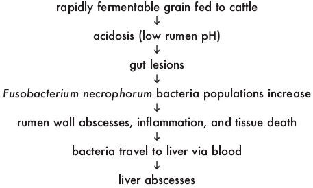
Both subacute and acute acidosis can lead to rumenitis (infection of the rumen wall). The low pH from acidosis creates lesions in the rumen wall. Damage to the rumen wall from sharp objects (such as wire or nails) predisposes the animal to abscess formation. When rumenitis develops, liver abscesses often follow. Bacteria (F. necrophorum, Actinomyces pyogenes, Bacteriodes spp.) from the rumen that cause liver abscesses enter the blood supply through ulcerative lesions, hairs, or foreign objects embedded in the rumen wall. These bacteria then travel via blood to the liver.
Liver abscesses are most often seen in feedlot cattle. Severe liver abscesses may reduce feed intake, weight gain, feed efficiency, and carcass yield. Abscessed livers are condemned at harvest, regardless of abscess severity. Liver condemnations from abscesses were observed in 13.5 per cent of fed cattle in the 2005 National Beef Quality Audit, resulting in about a 2 per cent reduction in carcass weight per head. Abscesses were the primary cause of liver condemnations.
Stages of Acidosis, Rumenitis, and Liver Abscess Development

Management Guidelines
When cattle are exposed to a high-concentrate diet too quickly, acidosis may result. Fluctuations in eating behavior are often observed. To reduce the incidence of acidosis, use a warm-up feeding period, introducing high-concentrate feeds gradually over three to four weeks. Keep at least 10 per cent roughage in the final diet to help moderate rumen pH. Forages and cottonseed hulls are both good roughage sources high in effective fiber.
While acidosis most often occurs during adaptation to concentrate-rich diets, chronic acidosis may continue into the feeding period. Feeding a combination of grains or feeding a dry grain with a high-moisture grain can reduce the risk of acidosis. Potential for acidosis is higher when feeding wheat than corn.
Processing grains less thoroughly and limiting the quantity of feed offered should reduce acidosis incidence but may also lower animal performance. Good bunk management where all feed is consumed before the next feeding may lessen daily fluctuations in feed intake and reduce acidosis risk.
Feeding ionophores (monensin or lasalocid) can help reduce the incidence of acidosis. Ionophores may reduce intake and help moderate concentrate intake when calves start on higher concentrate diets. Adding bicarbonate, fat up to 8 per cent of the diet, probiotics, virginiamycin, or thiamin to the diet or increasing protein in the diet may decrease acidosis risk.
Treatment for acidosis is similar to prevention efforts. Tylosin is effective in decreasing liver abscess incidence. Virginiamycin and chlorotetracycline also are used in addressing liver abscess problems.
Hardware Disease
Cause
Hardware disease is the common name for traumatic gastritis and traumatic reticulitis. It may occur when cattle consume sharp, heavy objects, such as nails or wire. These objects fall to the rumen floor and are swept into the reticulm by muscle contractions. Cattle may ingest these objects and never have hardware disease, or muscle contractions may cause these sharp objects to puncture the reticulum wall, diaphragm, and heart sac. Forceful abdominal movement during calving may force a sharp object through the reticulum wall. This leads to severe damage to and infection of the abdominal cavity, heart sac, or lungs.
Clinical Signs
Signs of hardware disease vary, depending on where the puncture occurs. Loss of appetite, depression, reluctance to move, arched back, and indications of pain are common signs. The animal may grunt when forced to walk. Recurring bloat may be noticed on the upper left side, with fluid accumulation on the lower right. If the heart sac has been punctured, fluid may accumulate around the heart and in the brisket. Fatal infection can occur if the object penetrates close to the heart.
Management Guidelines
Manage cattle so they can not ingest heavy, sharp objects. Keep pastures, paddocks, and feed bunks free of wire, nails, fencing staples, and other sharp objects (even heavy plastic items) that could be swallowed. Debris from structures and equipment may appear in areas where cattle graze after high winds. Deteriorating steel-belted tires also pose a risk.
Place magnets on feeding equipment to catch some of the metal objects in feed. An intraruminal magnet can be inserted into the rumen to trap metal fragments. Ingested metal is drawn to the magnet instead of working its way through the stomach wall. The magnet eventually “fills up” if enough metal is ingested. Administer a second magnet if signs of hardware disease persist. Magnets are relatively inexpensive, particularly when compared to the cost of surgery.
It is often difficult to diagnose hardware disease. Seek veterinary advice for suspect cases. Administer an intraruminal magnet when hardware disease can not be ruled out. Confining cattle and limiting feed intake may allow puncture sites to heal in less serious cases. If infection is suspected, administer a broadspectrum antibiotic.
The severity of infection and how long the condition has been present affect the animal’s treatment outlook. Cattle with extensive infection in the heart or abdomen have a very poor prognosis. Even with attempts to encourage feed consumption, these animals often die of starvation. Sometimes only surgical removal of the object works. Controlling infection is important after the object is removed for successful recovery. Surgery is often not a cost-effective option unless the cattle affected are very valuable.
Conclusions
Knowledgeable beef cattle producers can reduce or eliminate risk factors for bloat, acidosis, and hardware disease. Watch cattle for signs of nutritional disorders to facilitate early intervention and treatment. Solicit veterinary assistance when developing and implementing treatment programs for suspect cases of nutritional disorders. For more information on beef cattle nutritional disorders, contact an office of the Mississippi State University Extension Service.
References
Ball, D. M., C. S. Hoveland, and G. D. Lacefield. 2002. Southern Forages. 3rd ed. Potash and Phosphate Institute and Foundation for Agronomic Research. Norcross, GA.
National Cattlemen’s Beef Association. 2005. National Beef Quality Audit. Centennial, CO.
Owens, F. N., D. S. Secrist, W. J. Hill, and D. R. Gill. 1998. Acidosis in Cattle: A Review. J. Anim. Sci. 76:275–286.
U. S. Department of Agriculture. May 2007. National Cattle and Calves Nonpredator Death Loss in the United States, 2005. National Animal Health Monitoring System. Washington, D. C.
Vasconcelos, J. T., and M. L. Galyean. 2008. ASAS Centennial Paper: Contributions in the Journal of Animal Science to understanding cattle metabolic and digestive disorders. J. Anim. Sci. 2008:jas.2008-0854v1.
March 2009


