

Liver Fluke Control in Beef Cattle<sup>1</sup>
By M.B. Irsik, Charles Courtney III, and Ed Richey2, University of Florida, IFAS Extension. The common liver fluke (Fasciola hepatica) is recognized as one of the most damaging parasites in Florida cattle.The liver fluke is a problem in the Gulf Coast states and the Pacific Northwest; in Florida, most infected cattle are found grazing low-lying pastures in the peninsula, south and east of the Suwannee River. Few, if any, liver flukes are found in the panhandle of Florida.
Life Cycle
The adult liver fluke resides in the bile ducts of the animal's liver. The adult liver flukes produce eggs which are carried with bile to the gut and are then passed in the feces (see Figure 1).
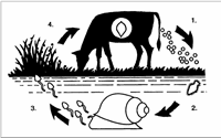 |
| Figure 1. Life Cycle Fasciola hepatica |
In the fecal pat, a ciliated larva called a miracidium develops inside the liver fluke egg. Complete development of the miracidium requires between 10 days to several months depending upon the temperature and moisture available. Development of the miracidium is known to occur at temperatures between 50ºF and 86ºF, ideal Florida weather. When a fluke egg is exposed to sunlight a developed, free-swimming, ciliated miracidium hatches. After hatching, the miracidium (see Figure 2) must find and penetrate a suitable snail host within a few hours or die. Inside the snail host, the miracidium multiplies and transforms into many tadpole-like cercariae which exit the snail. The penetration of a snail by a single miracidium can result in the production of hundreds of cercariae exiting the snail (see Figure 3).
The development of cercariae within the snail requires 5 to 7 weeks under optimal conditions (if, however, the miracidium infected snail begins estivation to escape the hot dry weather, recent evidence indicates that the infected snail will not survive the estivation period). After exiting the snail the tadpole-like cercaria attaches to vegetation, secretes a protective cyst covering, and completes its development into an encysted metacercaria. Each encysted metacercaria contains a fully developed immature fluke and is the infective stage of the parasite. The length of time that metacercariae survive on pasture is dependent on both moisture and temperature. A minimum of 70% relative humidity is considered necessary for prolonged survival of metacercariae. Metacercariae can be killed within 2 days when exposed to direct sunlight in temperatures of 98.6° to 105°F.
Cattle become infected by ingesting the metacercariae attached to forage or by drinking water contaminated with metacercariae attached to soil particles or vegetative debris (see Figure 4).
Once ingested and after reaching the small intestine, the infective fluke larva is released from the metacercaria, penetrates the wall of the small intestine and enters the abdominal cavity. After migrating through the abdominal cavity for approximately a week, the fluke larva penetrates the liver capsule and migrates slowly through the liver for 6 to 8 weeks. Finally, the fluke larva enters the bile duct where it matures and begins to produce eggs that are carried in the bile to the gut, thus completing the liver fluke cycle. Completion of the entire life cycle, from the time a fluke egg is shed onto the pasture until a newly infected animal reinfects the pasture with the next generation of fluke eggs, requires 16 to 24 weeks. Liver fluke transmission is the direct result of interactions between the parasite, the environment and the intermediate host. Cattle are usually infected with numerous liver flukes at any one time rather than a single liver fluke. Once ingested and after reaching the small intestine, the infective fluke larva is released from the metacercaria, penetrates the wall of the small intestine and enters the abdominal cavity. After migrating through the abdominal cavity for about a week, the fluke larva penetrates the liver capsule and migrates through the liver for 6 to 8 weeks. Finally, the fluke larva enters the bile duct where it matures and begins to produce eggs that are carried in the bile to the gut, thus completing the liver fluke cycle. Completion of the entire life cycle, from the time a fluke egg is shed onto the pasture until a newly infected animal reinfects the pasture with the next generation of fluke eggs, requires 16 to 24 weeks. Liver fluke transmission is the direct result of interactions between the parasite, the environment and the intermediate host. Cattle are usually infected with numerous liver flukes at any one time rather than a single liver fluke.
Damage to the Animal and Industry
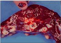 |
| Figure 2. Liver Damage Caused by Flukes |
Scar tissue replacement of damaged liver tissue caused by the migration of F. hepatica larvae through the liver is very pronounced. In the bile ducts, the adult flukes produce a mechanical irritation that causes an inflammation of the ducts that leads to a thickening and, in severe cases, calcification of the duct wall which can eventually result in blockage of the bile duct. Livers exhibiting any of these characteristics (scarring or duct damage) are condemned at slaughter (see Figure 2).
The condemnation of damaged livers at slaughter and the losses in beef production associated with fluke infections are economically significant. It has been estimated that the Florida beef industry loses $10 million each year due to liver fluke infections. Fascioliasis (liver fluke disease) is associated with reduced fertility of the brood cow herd, lighter calves at weaning, slower growth of replacement heifers, higher culling rates in cow herds and lighter weight cull cows. In the dairy industry, a reduction in milk production (10%) occurs in fluke-infected herds; in addition, fluke infected replacement heifers take longer to reach breeding age. Although cattle seldom die of fluke infection, an occasional secondary Clostridium haemolyticum infection in damaged livers may cause death in animals not properly vaccinated against "red water" disease.
Life Cycle of the Intermediate Host
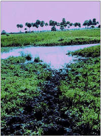 |
| Figure 3. Cattle Trail Crossing Drainage Ditch |
In Florida, the lymnaeid snails Pseudosuccinea columella and Fossaria cubensis serve as the intermediate hosts of the liver fluke with F. cubensis being the more prevalent and the most important intermediate host. Lymnaeid snails are semi-aquatic/amphibious snails that prefer a wet, muddy environment. Large populations of lymnaeid are almost never found in areas covered with vegetation or areas that are permanently flooded. Lymnaeid snails frequently can be observed below the water surface during the cooler portions of the year when surface soil and water temperatures are warm. Lymnaeid snails are almost always on the mud surface if open mud surface is available or attached to vegetation above the water line if the area is temporarily flooded.
An excellent environment for the snail is established where frequently used cattle trails cross drainage ditches or lead to watering ponds (Figure 3). The continuous trampling of the wet soil kills the vegetation and the crossings become open mud areas that provide an excellent environment for the lymnaeid snails to inhabit. If cattle are removed from the pasture and the crossings are not used, grass and weed growth will soon cover the open muddy areas, thus destroying the environment that lymnaeid snails prefer. Lymnaeid snails rarely inhabit pastures free of open mud areas. If lymnaeid snails are found in dry pastures they were probably brought into the area by a recent flood and will not survive.
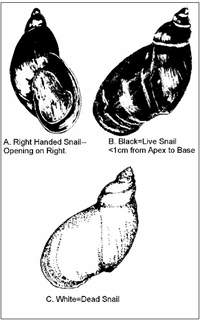 |
| Figure 4. Lymnaeid Snails Pseudosuccinea columnella and Fossaria cubensis |
Lymnaeid snails are small (<1cm), "right handed" snails that can be recognized by placing the snail on its back with the apex or peak of the snail up and the opening of the shell (orifice) facing you. The opening or orifice is on the right side of a "right handed" snail (Figure 4A). The shell of a living lymnaeid snail is black in color (Figure 4B). When the snail dies, the shell becomes white (Figure 4C).
Hot dry weather causes snails to dehydrate and die; however, lymnaeid snails have the ability to survive adverse times (hot dry weather) by burrowing into the mud and entering a state of reduced metabolic activity called estivation (similar to hibernation). Snails that survive the hot dry periods of the year by estivation emerge when the environmental conditions improve in the fall season. Lymnaeid snails are hermaphroditic; therefore, the entire mature population is capable of producing fertile eggs. Some lymnaeid snails reach maturity and begin to produce eggs within 14 days of being hatched. Once lymnaeid snails begin to lay eggs, they will continue to do so for the rest of their lives (3-7 months), ceasing only while in estivation. One mature snail can easily produce 5,000 eggs in a lifetime. This allows for tremendous increases in snail populations over a relatively short period of time whenever environmental conditions are favorable. Favorable environmental conditions for the growth and reproduction of lymnaeid snails generally occur in Florida from late September through June in the northern part of Florida and late October through May in southern Florida.
A problem in making broad statements such as this is that snails will emerge from estivation at slightly different times each autumn depending upon interactions of climatic and environmental variables that differ each year. Weather is the key to triggering the beginning of snail estivation; in Florida hot and dry weather will trigger the beginning of snail estivation. However, we also find very few lymnaeid snails during Florida's wet hot summers. Cooler wet weather triggers the emergence of the snail from estivation. If snail habitats remain dry in the late summer and early fall, emergence of the snails from estivation will be delayed. Any change (early or late) from the normal beginning or ending of the hot dry season causes a respective change in the beginning or ending of snail estivation.
Diagnosis of Liver Fluke Infection
Microscopic examination of cattle feces for the presence of liver fluke eggs still remains the most popular method of diagnosing liver fluke infections in cattle herds. However, this technique is labor intensive (requiring 20-30 minutes per sample) and may result in missed diagnosis because of low egg output in infected cattle, poor sedimentation properties of the fluke egg, and excessive fecal debris on the microscopic slide that obscures the fluke eggs during examination. A modification of the fecal examination technique which includes the use of a two-sieve filtering system (Flukefinder®, by Visual Difference, Moscow, ID) improves the speed of the process and fluke egg detection.
Several factors need to be recognized when interpreting fecal examinations for the detection of liver flukes. Liver flukes are generally not evenly-dispersed within a herd of cattle; a small percentage of cattle in a herd will carry the greatest fluke loads and therefore shed the most fluke eggs. In addition, fluke infected cattle shed relatively few eggs (less than 5 eggs per gram of feces, even in heavily infected herds); therefore a minimum of 10 samples should be examined before any diagnosis of fluke infection or lack of infection is made on any cattle herd. However, the best diagnostic tool would simply be to examine the livers of your cull cows while they are being slaughtered (Figure 2). Arrangements can and should be made for you or your veterinarian to examine the livers at the time of slaughter.
Control of the Liver Fluke
Treatment of beef cattle in Florida during late summer, ideally between August 15 and September 1, will eliminate flukes acquired during spring & early summer. This is the weak point in the liver fluke's life cycle - flukes survive the summer in Florida only as adult flukes in the livers of cattle. Liver fluke eggs shed in feces during the summer are usually killed by the combination of summer heat and rainfall, metacercariae encysted on vegetation are quickly killed by summer's heat and are not replaced after snails enter estivation, and fluke-infected snails seldom survive estivation. Fluke treatment between August 15 and September 1 kills the adult flukes. This prevents cattle from shedding fluke eggs onto pasture when snails emerge from summer estivation with the onset of wet, cool weather and thereby breaks the life cycle of the liver fluke.
At the present, flukicidal products marketed on the U.S. market do not reliably kill juvenile flukes (larval flukes not yet in the bile ducts of cattle, i.e. < 8 weeks old). Clorsulon (Curatrem®)and albendazole (Valbazen®) are the only approved flukicides for use in cattle in the United States. A full dose (7 mg/kg orally) of Clorsulon is highly effective against mature (egg laying, i.e. > 12 weeks old) and immature flukes (8-12 weeks old) but, unlike albendazole, has no activity against nematodes. However, clorsulon is compatible with concurrent administration of benzimidazoles, levamisole, and ivermectin. At a reduced dose (3.5 mg/kg) clorsulon is effective only against mature flukes. This dose should only be used at times of the year (i.e. late summer) when you are certain no immature flukes are present. Clorsulon also is available in combination with ivermectin (Ivomec Plus®). This injectable formulation delivers 2 mg/kg clorsulon, biologically equivalent to the 3.5 mg/kg "half dose" of oral clorsulon. The efficacy of albendazole is similar to that of the reduced dose of clorsulon and is only effective against mature (>12 weeks old) flukes. Because of the lack of efficacy of the flukicides upon juvenile flukes, spring treatments, when all ages of flukes are present in infected cattle, will only temporarily reduce the fluke numbers. Spring fluke treatments can be helpful to reduce fluke exposure to calves and young cattle but by no means can a spring treatment be expected to provide the fluke control required in infected herds in Florida. Albendazole should not be administered to any female cattle during the first 45 days of pregnancy. Clorsulon and albendazole are not approved for use in adult dairy cattle.
In extremely wet years, or on ranches with severe fluke problems, a second treatment at spring roundup may be cost effective. Autumn hurricanes may bring cool, wet weather early in the year (as occurred in autumn of 1985), allowing snails to emerge from estivation 1-2 months earlier. Under these circumstances some fluke-infected snails may survive estivation and fluke transmission to cattle may begin as early as October. Treatment of adult dairy cows at calving and again at drying off (when approved flukicides become available for that use) may give acceptable fluke control without necessitating the disposal of otherwise saleable milk, although it would not break the life cycle as effectively as the seasonal treatment scheme recommended above for beef cattle. This seasonal scheme is not appropriate for adult dairy cows since few dairymen will want to discard perfectly good milk.
Snail control will reduce fluke infestation on ranches; however, no products that kill snails (molluscicides) are approved by the Environmental Protection Agency because they kill too many other non-target aquatic organisms. Therefore, "snail sprays" such as copper sulfate that may have been referred to in older Extension publications are not allowed. Ditching and draining pastures will help reduce snail habitat, but in Florida the Water Management Districts will seldom grant permission to drain wetlands. By virtue of its constant temperature and near neutral pH, artesian ("flowing") well runoff may support snail populations year round and be should minimized to the greatest extent possible. Where such runoff cannot be eliminated, substituting a bare sand bottom in place of organic muds will greatly reduce snail populations by eliminating their food supplies.
Cattle in Florida occasionally are infected with the deer fluke Fascioloides magna. Little damage appears to be done other than the occasional condemned liver in cull cows, and no control measures appear justified in Florida at this time. Likewise Florida cattle commonly are infected with rumen flukes (paramphistomes) which are apparently harmless. However, in other parts of the U.S. the deer fluke and rumen flukes have caused clinical disease and/or production losses in cattle.
Footnotes
1 This document is VM120, one of a series of the Veterinary Medicine-Large Animal Clinical Sciences Department, Florida Cooperative Extension Service, Institute of Food and Agricultural Sciences, University of Florida. Original publication date April 22, 2002, revised July, 2007. Visit the EDIS Web Site at http://edis.ifas.ufl.edu.
2 M.B Irsik, Assistant Professor, Charles H. Courtney, Professor and Associate Dean for Research, and Ed Richey, Extension veterinarian, retired, Department of Large Animal Clinical Sciences, College of Veterinary Medicine, Cooperative Extension Service, Institute of Food and Agricultural Sciences, University of Florida, Gainesville, 32611.
Further Reading
| - | Find out more information on Liver Fluke by clicking here. |
April 2008


